Illustration: Male Human Skeleton Femur Muscle Anatomy. 3D Render
ID 275692061 © Paihub | Megapixl.com
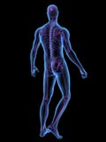
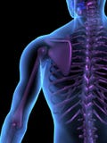
The femur, also known as the thighbone, is the largest and strongest bone in the human body. It is located in the upper leg, extending from the hip to the knee joint. The femur is surrounded by several muscles that attach to it, allowing for movement of the leg.The muscles that attach to the femur include:Quadriceps femoris muscle: This muscle group includes the rectus femoris, vastus lateralis, vastus medialis, and vastus intermedius muscles. These muscles originate from the pelvis and femur and attach to the patella and tibia via the patellar tendon. They are responsible for extending the knee joint and flexing the hip joint.Hamstring muscles: The hamstring muscle group includes the biceps femoris, semimembranosus, and semitendinosus muscles. These muscles originate from the ischial tuberosity of the pelvis and attach to the tibia and fibula bones of the lower leg. They are responsible for flexing the knee joint and extending the hip joint.Gluteus maximus muscle: This muscle originates from the ilium bone of the pelvis and attaches to the femur bone. It is responsible for extending the hip joint and rotating the thigh laterally.
CATEGORIES
Sharing is not just caring, it's also about giving credit - add this image to your page and give credit to the talented photographer who captured it.:
KEYWORDS
skeleton illustrationqua illustrationquad quaanatomy muscle quadriceps femoris large composed distinct heads rectus superficial muscles middle originates male human anatomy triceps render brachii commonly upper arm humerus radial dorsri latissimus dorsi triangular located movements including shoulder spinous thoracic vertebrae thoracolumbar attaches intertubercular groove humanmuscle skeletion femour femur thighbone largest hip knee surrounded movement lateralis medialis intermedius originate pelvis patella tibia patellar tendon responsible flexing biceps
More images on Dreamstime


Similar Images


More images by the same author


1 week free trial. Any size, for free
Renews at $25/month, cancel or upgrade at any time
Click here for your free trial!240,983,383 images | 1,214,938 contributors | 53,325,448 total users